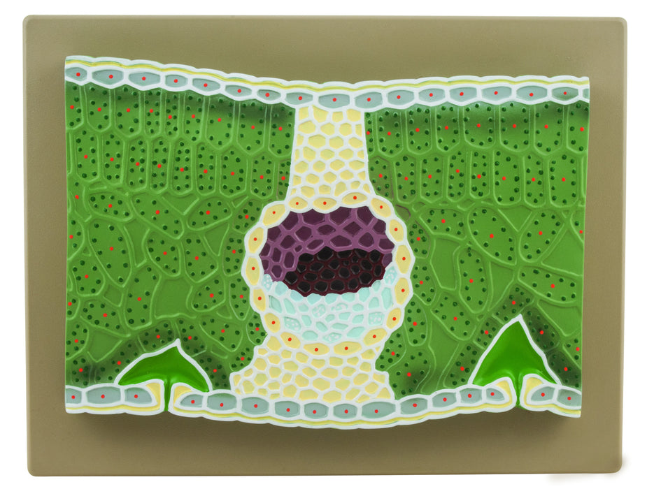
Dicot Leaf Model, on Base – 11″ – Greatly Magnified – Eisco Labs
The detailed 3D rendering of a dicot leaf section, which is greatly magnified, is ideal for studying the structure and function of a dicot leaf. Structures featured include: cuticle, upper epidermis, hypodermis (collenchyma), pallisade parenchyma, spongy parenchyma, air spaces, chloroplasts, bundle sheath, sclerenchyma, xylem, phloem, lower epidermis, guard cells, and stomata. These key features are colored and numbered for comparison with the key card that is included. This model provides a visually and kinesthetically effective method for studying the structure and function of the various structures of a dicot leaf.


Reviews
There are no reviews yet.