This blog is on the Principle, Price, and Result Samples of Scanning Electron Microscope(SEM)
The types of signals produced by a Scanning Electron Microscope(SEM) include secondary electrons (SE), back-scattered electrons (BSE), characteristic X-rays, light (cathodoluminescence) (CL), specimen current, and transmitted electrons.
Secondary electron detectors are standard equipment in all SEMs, but it is rare that a single machine
would have detectors for all possible signals. The signals result from interactions of the electron
beam with atoms at or near the surface of the sample. In the most common or standard detection
mode, secondary electron imaging or SEI, the Scanning Electron Microscope(SEM) can produce very high-resolution images of a sample surface, revealing details less than 1 nm in size.
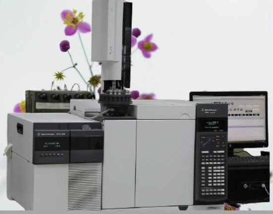
Due to the very narrow electron beam, SEM micrographs have a large depth of field yielding a characteristic three-dimensional appearance useful for understanding the surface structure of a sample. This is exemplified by the micrograph of pollen shown above.
A wide range of magnification is possible, from about 10 times (about equivalent to that of a powerful hand lens) to more than 500,000 times, about 250 times the magnification limit of the best light microscopes.
Back-scattered electrons (BSE) are beam electrons that are reflected from the sample by elastic
scattering. BSE is often used in analytical Scanning Electron Microscope(SEM) along with the spectra made from the characteristic X-rays because the intensity of the BSE signal is strongly related to the atomic number (Z) of the specimen. BSE images can provide information about the distribution of different elements in the sample. For the same reason, BSE imaging can colloidal gold immune labels of 5 or 10 nm diameter, which would otherwise be difficult or impossible to detect in secondary electron images in biological specimens.
Characteristics X-rays are emitted when the electron beam removes an inner shell electron from the
sample, causing a higher-energy electron to fill the shell and release energy. These characteristics Xrays are used to identify the composition and measure the abundance of elements in the sample.
Principle of Scanning Electron Microscope (SEM)
All samples must be of an appropriate size to fit in the specimen chamber and are generally mounted
rigidly on a specimen holder called a specimen stub. Several models of Scanning Electron Microscope(SEM) can examine any part of a 6-inch (15 cm) semiconductor wafer, and some can tilt an object of that size to 450
.
Samples are coated with a platinum coating of electrically conducting material, deposited on the
sample either by low-vacuum sputter coating or by high-vacuum evaporation. SEM instruments
place the specimen in a relatively high-pressure chamber where the working distance is short and the
electron optical column is differentially pumped to keep the vacuum adequately low at the electron gun.
The high-pressure region around the sample in the ESEM neutralizes charge and provides an
amplification of the secondary electron signal. Low-voltage Scanning Electron Microscope(SEM) is typically conducted in a FEGSEM because the field emission guns (FEG) is capable of producing high primary electron brightness and small spot size even at low accelerating potentials.
Embedding in a resin with further polishing to a mirror-like finish can be used for both biological and
materials specimens when imaging in backscattered electrons or when doing quantitative X-rays
microanalysis.
Results from Our Scanning Electron Microscope (SEM) Units
Below is a link to sample results from our Scanning Electron Microscope analysis. Our SEM unit has been argued as one of the best in Nigeria and we think that is correct.
Prices of our SEM, SEM-EDS Analytical Units
Currently, we have three units for Our Scanning Electron Microscope (SEM). Unit one is considered our best of all.
End note:
This blog is on the Principle, Price, and Result Samples of Scanning Electron Microscope(SEM). The ideas expressed is a revision of the Wikipedia archive on the subject. All questions and comments shoule be directed to research@allschoolabs.com. Thank you
 No products in the cart.